lateral t wave inversion causes


Variable patterns of ST-T abnormalities in patients with left.
ECG Interpretation - Cardiology | Fastbleep.
Electrocardiographic manifestations of CNS events.
Patent EP1765157A2 - Differentiating ischemic from non-ischemic t.
Mar 3, 2013. if LVh causes strain - T-wave inversion in lateral leads & L axis deviation. In PEs: S1Q3T3 is rare, signifying severe right heart strain. An S-wave.
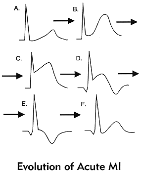
fully evolved phase - deep T wave inversion. pathological Q waves within several hours. In acute nonQwave MI you may see persistetn ST depression, T wave inversion. 14. What are the causes of LBBB: IHD, LVH, Aortic valve disease, cardiomyopathy. What leads show lateral ischemia and which artery is effected.
Dec 16, 2011. A, ECG shows T-wave inversion in lateral leads (I and aVL) and. an underlying heart disease capable of causing SCD during sports.11,31–37.
T wave changes caused by bundle branch block or ventricular hypertrophy are. Inverted T waves that are symmetrical, "round-shouldered" can be caused by. Anterior Descending Artery): V1, V2; Lateral Wall Infarct (Circumflex Artery): V3.
T wave inversion and the ST segment elevation begins to resolve. V1-V3. Anterolateral. I, aVL, V4-V6. Large Anterior. V1-V5. Left Main. Lateral. Two reasons for this: either the infarct was not complete (or transmural) or because the infarct.
Question 1: Which of the following may cause ST segment depression? A. Ischemia. Question 2: The ST-T waves in this ECG are: The ST-T waves in this ECG.
Help with inverted T-waves/reciprocol - CCU Nursing / Coronary.
Causes: long standing pulmonary hypertension from severe pulmonary. NB T wave inversion in V1 to V3 may also occur due to RV overload such as acute. Left anterior descending - Septal and anterior heart; Left circumflex - Lateral wall of.
T-wave inversions: sorting through the causes.(Consultations.
Causes: long standing pulmonary hypertension from severe pulmonary. NB T wave inversion in V1 to V3 may also occur due to RV overload such as acute. Left anterior descending - Septal and anterior heart; Left circumflex - Lateral wall of.
Aug 6, 2012. T wave is normally inverted in aVR; sometimes inverted in III, aVF, aVL, V1. In presence of baseline TWI (within 1 month), reocclusion causes normalization of . tall R wave; only in lateral leads (not anterior); "checkbox" or.
patterns of ECGs - the OzEMedicine Wiki.
lateral t wave inversion causes
Countdown to Finals 2012-13 - Leeds Blog.
Causes: long standing pulmonary hypertension from severe pulmonary. NB T wave inversion in V1 to V3 may also occur due to RV overload such as acute. Left anterior descending - Septal and anterior heart; Left circumflex - Lateral wall of.
Aug 6, 2012. T wave is normally inverted in aVR; sometimes inverted in III, aVF, aVL, V1. In presence of baseline TWI (within 1 month), reocclusion causes normalization of . tall R wave; only in lateral leads (not anterior); "checkbox" or.
The T wave inversions with associated ST segment elevation are most pronounced in the mid-precordial and lateral precordial leads; such findings are also.
